Evaluating Modern Deep Learning Architectures for Brain Tumor Detection
Abstract
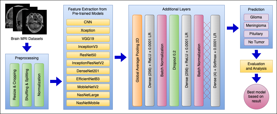
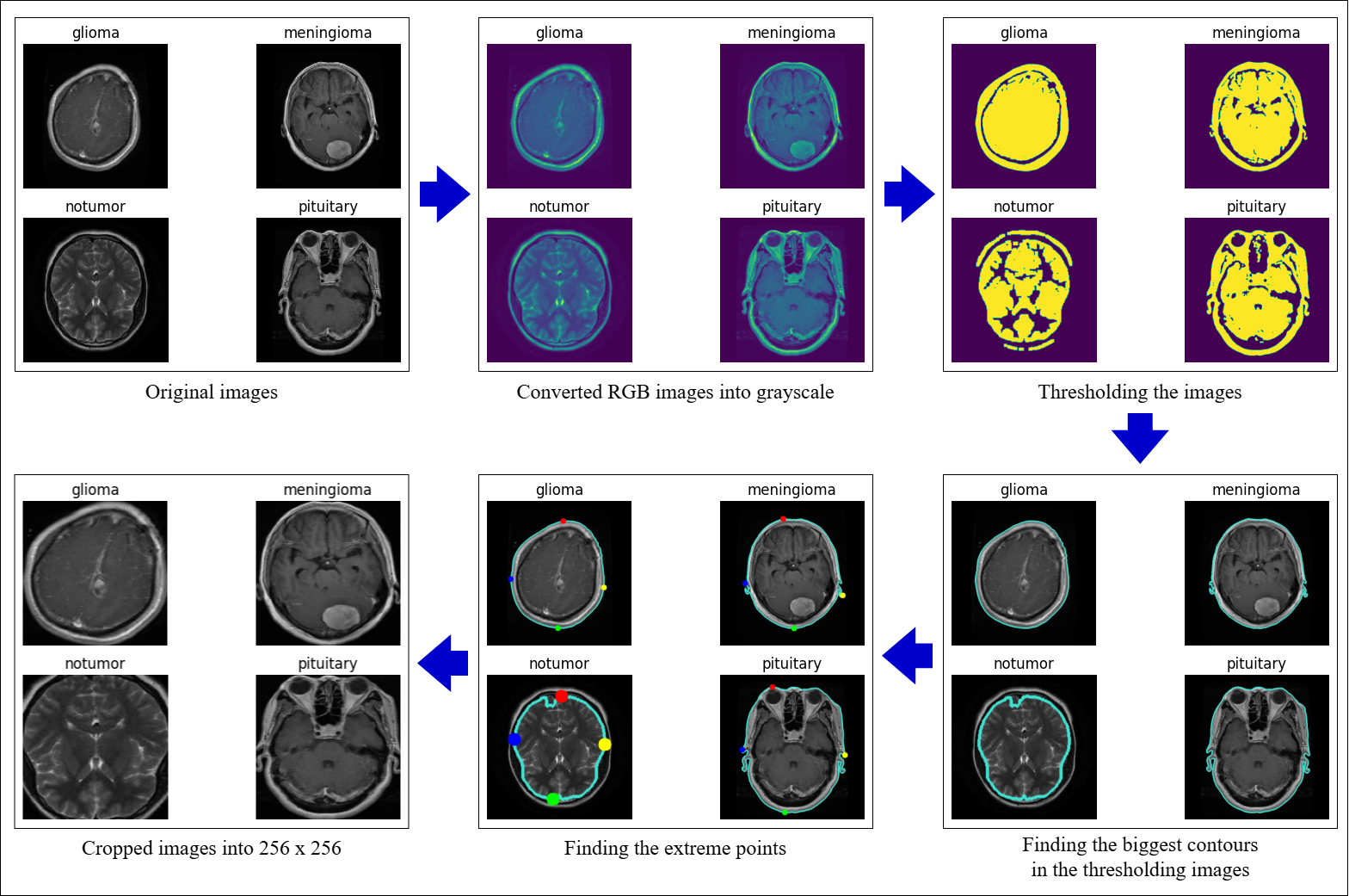
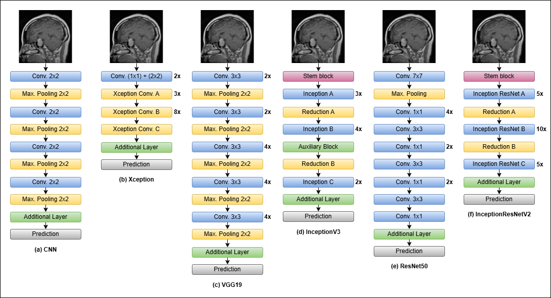
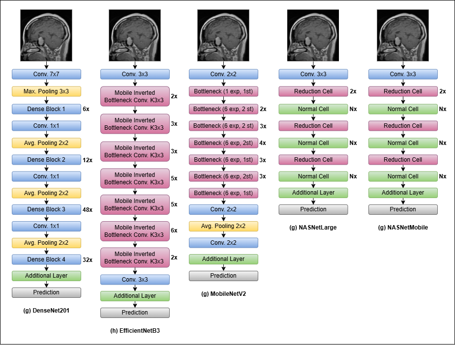
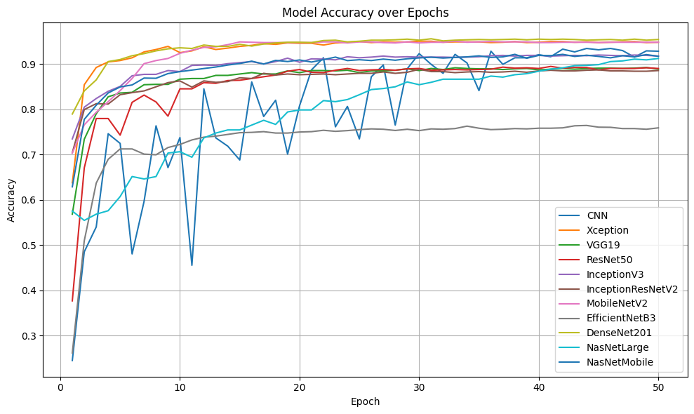
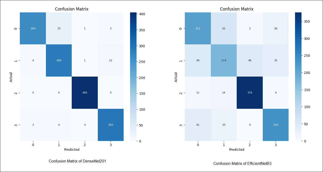
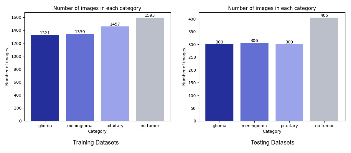
Brain tumors are among the most lethal types of cancer, with high mortality rates and aggressive progression. Early detection through imaging, particularly MRI, is crucial for improving survival rates. This study evaluates 11 deep learning models which has not been done in previous studies, including CNN, Xception, VGG19, InceptionV3, ResNet50, Inception-ResNetV2, DenseNet201, EfficientNetB3, MobileNetV2, NASNet-Large, and NASNetMobile, for their effectiveness in brain tumor detection using MRI scans. All models were trained and tested with identical preprocessing and hyperparameter settings to compare their performance. Adam optimizers were used to train each model with a learning rate of 0.0001, decay steps of 100, and a decay rate of 0.95 across 50 epochs. Dropout is used to reduce overfitting and speed up the learning process, with a rate of 0.2. L1 and L2 regularization are applied to the three dense layers to prevent overfitting and help the model learn better as it becomes more complex. Each dense layer uses the ReLU function, with 256 neurons in the first layer, 128 neurons in the second, and a softmax function in the final layer for classification. The final results show the DenseNet201 model achieves the highest accuracy of 95.65%, while the EfficientNetB3 model achieves the lowest accuracy of 75.89% on the testing data. These findings suggest that deep learning models like DenseNet201 show real potential for enhancing medical imaging diagnostics. Additional research on hybrid architectures, larger and clinical data validation, and fine-tuning are needed in order to improve the generalizability and accuracy of models.
Paper Information
- Category Computer Vision, Deep Learning, Data Science
- Publisher IEEE
- Publish date August 28, 2025
- Organization 2025 International Conference on Information Management and Technology
- View Paper